This is a case of altered mental status encountered in the GI ward in September 2019, detailed below.
An 80 y/r female was found to have liver cirrhosis complicated by ascites earlier this year and was referred to our hospital's gastroenterology outpatient clinic. Blood tests showed HBV and HCV were both negative. During the week of 8/11, the patient's consciousness gradually deteriorated, accompanied by nausea, vomiting, poor appetite, and constipation. Following an outpatient visit on 8/14, she was admitted on 8/15 for ascites tapping and lab testing.
At the bedside, the patient was assessed to have a GCS of E2V2M4. The patient appeared extremely emaciated overall, with almost no muscle, while presenting with abdominal distension that yielded a dull sound upon percussion. From her family's account and follow-up medical records, it was noted that her weight had dropped 15 Kg from March to the present.
In gastroenterology, the most common cause of altered mental status is Hepatic encephalopathy, but of course, other causes must be ruled out. Using the mnemonic A-E-I-O-U-T-I-P-S, considering the history, besides basic biochemistry, CBC/DC, we checked blood glucose, electrolytes, Ammonia, and Vein Gas. Although there was no fever, two sets of blood cultures were drawn to rule out infection under the VS attending physician's recommendation. It was also pointed out that regarding electrolytes, the main ones affecting consciousness are Na and Ca.
(A more senior VS mentioned he felt that checking routine Ammonia is meaningless because its blood concentration doesn't completely correlate with hepatic encephalopathy; even if you draw a value within the normal range, you cannot rule out hepatic encephalopathy.)
Because this patient arrived around 4 PM, when the hospital's lab value alert text message came, I was still in the hospital. Fearing the on-call physician's workload would be too heavy, I handled it myself, and ended up dealing with it until past 8 PM, so the impression is still very deep now.
Blood calcium was as high as 14.5 mg
Blood calcium was as high as 14.5 mg
Blood calcium was as high as 14.5 mg
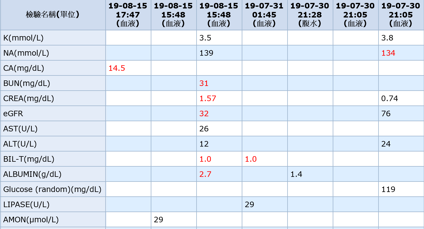
Well then, after a search, emergency symptom management mainly involves aggressive hydration for Dilution and giving calcium-lowering drugs. After inquiry, subcutaneous Calcitonin injection was chosen on the spot. An EKG also had to be done. Being in my first month as a PGY, I was on edge about everything. The sticking point was: ah, there's already ascites present, wouldn't giving hydration just put more fluid into the abdominal cavity? But finally, weighing the dangers of hypercalcemia, the ascites could only be considered a minor issue. Other significant Lab data included Anemia, Thrombocytopenia, Acute Kidney Injury, and Hypoalbuminemia.
Later during admission, after emergency Hydration and Calcitonin injection, the patient's blood calcium dropped, and her consciousness gradually cleared. The original symptoms of nausea, vomiting, and constipation also subsequently improved. But the underlying cause wasn't resolved; the hypercalcemia kept relapsing. In the lengthy hypercalcemia workup process, every week when the blood calcium spiked, Hydration had to be continued. In between, there were even episodes of pulmonary edema and hypokalemia. The patient's family and primary nurse expressed mental exhaustion, and I felt very guilty whenever explaining the disease to the family and primary nurse...
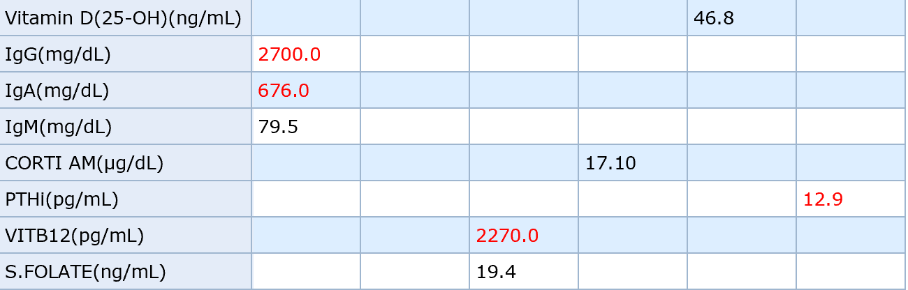
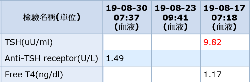
The Survey process was very tortuous. Synthesizing various guidelines, we first had these results: 1. PTHi was low. 2. Vitamin D 25(OH)D was in the normal range. Our hospital didn't have 1,25(OH)2D or PTHrP. After checking thyroid function, it was found to be Subclinical Hypothyroidism.
Tracing back the patient's history, the family clearly stated that the patient only started lying in bed after her consciousness altered; otherwise, she could usually walk with a walker and do chores herself. The family also mentioned there was no family history of hypercalcemia, nor was she taking calcium tablets. Additionally, she had visited a pain clinic for back and bone pain due to HIVD.
Synthesizing all this, the investigation launched mainly in three directions:
- Multiple myeloma or other bone marrow diseases
- Tuberculosis infection
- Cancer
Multiple Myeloma
Multiple myeloma was suspected because the patient actually met the symptoms of CRAB:
- C - Hypercalcemia
- R - Renal complications
- A - Anemia
- B - Bone disease
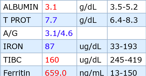
A reverse A/G ratio was observed in this patient, but this could appear in patients with either liver cirrhosis or Multiple myeloma. The former is due to low Albumin, while the latter is due to an abnormal increase in globulins. For this patient, it was hard to determine.
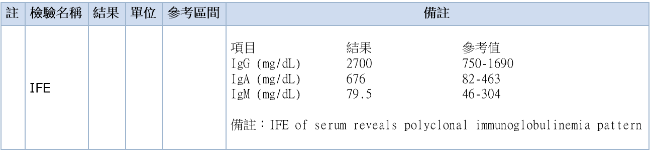
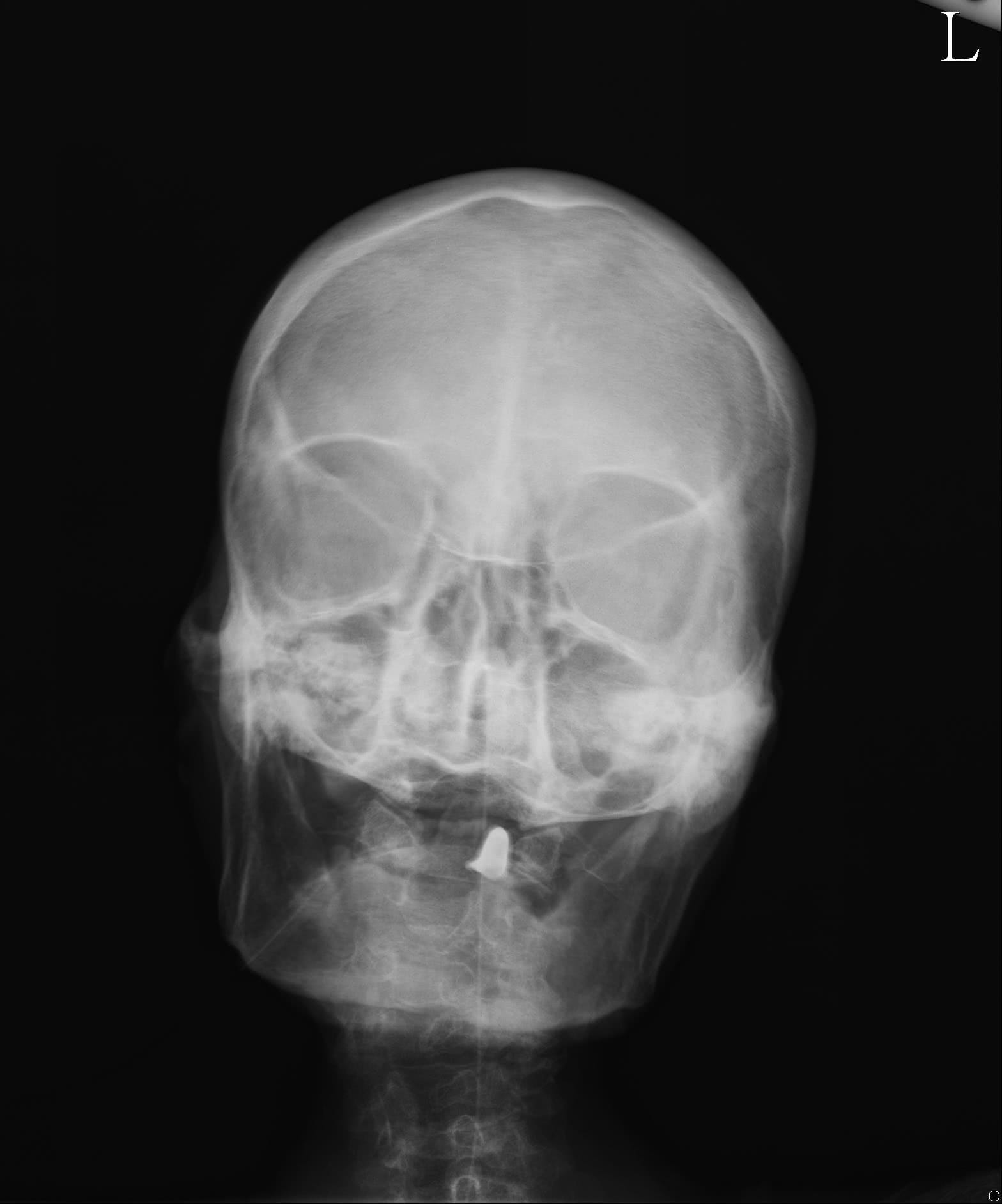
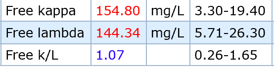
We checked IFE (Immunofixation Electrophoresis) and found increases in both IgA and IgG, presenting a polyclonal pattern, which is less like the monoclonal pattern of Multiple myeloma. An examination of the Skull X-ray did not reveal the osteolytic lesions of Multiple myeloma. Checking the kappa/lambda ratio, it was also normal.
Ascites
The patient's Chest X-ray did not show characteristics of pulmonary tuberculosis. Two sets of sputum left for Acid-fast stain were also negative; a third set was delayed at the turn of the month because the patient had no sputum. What raised suspicion was the abdominal ultrasound of the ascites.
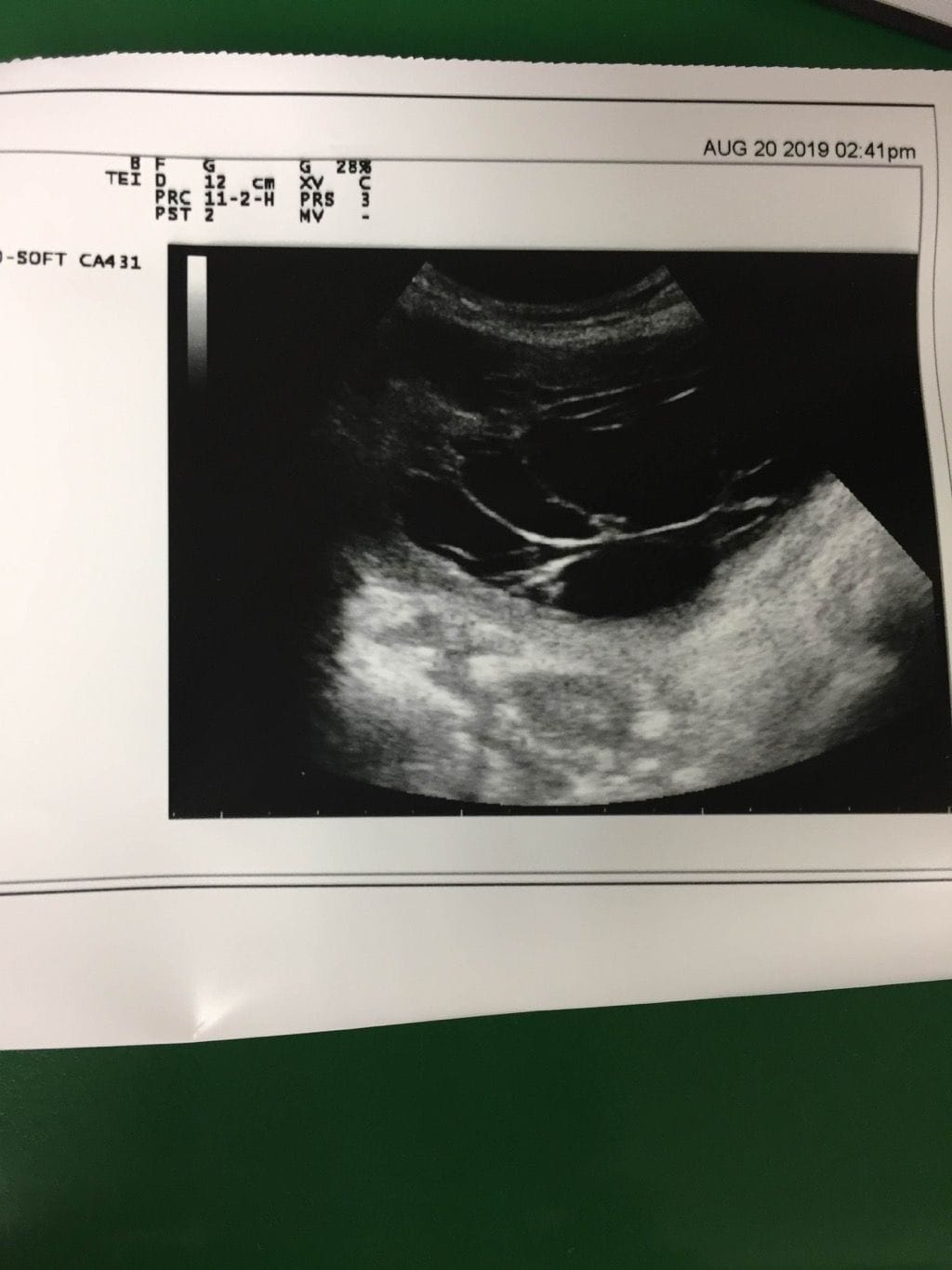
Under ultrasound, characteristics of loculated ascites could be seen. After consulting a senior resident, we had to suspect peritonitis or peritoneal carcinomatosis infection. It could also present this way if Abdominal tapping had been done multiple times to drain ascites, but this was the patient's first time having ascites drained.
Hmm, so the ascites was sent to test a bunch of things.

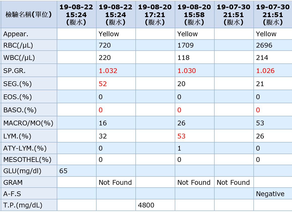
At least the ascites Data didn't look like an SBP infection, but the strange point was the very high Total protein. If calculating the SAAG, it would definitely be less than 1.1. The disease could be TB peritonitis, peritoneal carcinomatosis, pancreatic ascites, or nephrotic syndrome. Among these, TB peritonitis and peritoneal carcinomatosis were exactly the possible causes we suspected for the hypercalcemia. After ruling out others from the Data, we sent the ascites again for TB-PCR and Cell cytology (to look for malignancy), and the results were all negative...
Possibility of Cancer?
Losing a lot of weight in a short period, combined with hypercalcemia, definitely makes the possibility of Cancer very high. But an awkward point for the patient was that she had just had an abdominal CT scan at the end of July when coming to the ER for fever and abdominal pain. If we were to do it again at the end of August, there might be issues with National Health Insurance deductions and radiation exposure. At that time, the abdominal CT did not show lesions suspicious of Cancer. The earlier pathology report of the ascites was also negative. Head and neck as well as breast Physical Examinations did not show obvious mass lesions. The only thing not done was a chest CT...
Latest Progress Before the Month Turnover
Later, after the attending physician coordinated with the family, they decided to directly do a whole-body CT scan and a bone scan. There might also have been a factor of the family requesting less torment. However, a glance at the report seemed to show no obvious lesions in the chest; the main issue was still the abdominal ascites over there. I will continue to follow up when I have time later.